

New Techniques in Human Distal Convoluted Cell (hDCT) Culture
Kidney diseases and blood pressure cause >15 million death per year globally. The kidney’s distal convoluted tubules (DCT) were recently discovered to control blood pressure in response to dietary salt, however, current cell models poorly replicate human physiology for further study.
I aim to optimise a recently developed protocol for isolating and immortalizing primary DCT cells from healthy volunteers, instead, using clinically collected urinary samples from patient with renal tubular disorders and/or hypertension. These cells will be pivotal in the development of disease-specific models for research. Additionally, both immortalised and primary hDCT will be cultured on an “organ-on-a-chip” system to create 3D tubular-like structures that better emulate the real-life tissue architecture and physiological responses. The tubules can then be grown alongside artificial blood vessels to investigate tubule-capillary interactions. Unlike 2D models, this system can be perfused at different flow rates for functional experiments and pharmacological testing.
The ultimate goal of this project is to bring genuine personalised medicine to the diagnosis and treatment of tubular diseases and hypertension. Using this approach, we can tailor individual treatment plans to maximise patient benefit in a completely non-invasive, patient-centered manner using their own urine- and blood-derived cells.


SLC12A3 Biology in Health and Disease
High blood pressure, or hypertension (HT), is the leading risk factor for premature death worldwide. However, the underlying cause of HT remains unclear. By studying individuals with rare genetic disorders we hope to gain new insights into the causes of HT in the general population. The role of one particular kidney protein, the salt transporter SLC12A3 (produced from a gene with the same name), in the regulation of human blood pressure is especially interesting to our group. Elizabeth’s PhD project has two main focuses: First, she is exploring a patient who has low blood pressure and looks like she has a rare kidney disorder, Gitelman syndrome, but without a mutation in SLC12A3 which we expected to find. She is using a variety of laboratory techniques to explore the underlying cause of the disease in this patient. Second, she is looking at the general population (by using data in the UK Biobank), to see how individuals with changes in SLC12A3 are impacted through their lives. She will use complex statistical approaches, known as bioinformatics, to explore those features (such as blood pressure, blood results, heart shape and function) associated with mutations in SLC12A3 and other kidney protein genes.
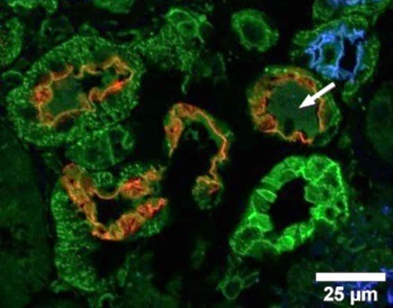

3D segmentation of the renal tubular structure from the optical cleared sample
Gold-standard diagnosis evaluates the nephron's patho-characteristic via kidney biopsy, yet the structural complexity of the intertwined tubules restricts the accuracy and efficiency. Here we want to propose a workflow which joins optical clearing and artificial intelligence to improve the accuracy and accelerate the process by automation substantially.
Optical clearing is an advanced tissue preparation protocol which turns the sample transparent, allowing us to image it from multiple directions without destroying its physical shape while increasing the resolution of the final 3D image. With machine learning, individual structural component is auto segmented for assessment, including length and volume.
We can then precisely map the interior tubular configuration to its 3D image, and measurements such as the cross-section area, length and volume of the tubular can be easily retrieved. Namely, a sample can be transformed into a glass LEGO so that every piece is dismountable for measurement automatically.
This workflow could support clinicians in generating appropriate diagnoses using precise sample analysis. In research, 3D spatial characterization of immunofluorescence-stained tissue provides crucial insights into the location and function of genes. This novel method is highly translational that can be migrated to other tubular organs, such as the lung, heart, and brain, benefiting the broader community with an easy adaptation of code.

Investigating the renal reabsorption of inorganic nitrate and its effect on blood pressure
Hypertension is the leading risk factor for premature death globally, causing a significant burden of morbidity, such as heart attacks, strokes, and renal failure. To inform dietary and pharmacological therapies, it is essential to identify the mechanisms that control blood pressure.
The role of dietary inorganic nitrate, found abundantly in green-leafy vegetables, is increasingly acknowledged for its ability to regulate blood pressure. Circulating nitrate is freely filtered at the glomerulus, but undergoes tubular reabsorption, which is the major factor contributing to its 6-hour half-life in the plasma. Data suggest that nitrate reabsorption occurs downstream of the loop of Henle, potentially in the collecting duct.
The role of Pendrin (SLC26A4), a chloride/bicarbonate exchanger in β-intercalated cells of the collecting duct, has generated interest in the context of hypertension, as it appears to work in conjunction with another transporter to absorb NaCl in the collecting duct.
To understand the role of pendrin in hypertension, an in vitro 3D model of the collecting duct will be developed using an organ-on-chip system. This system will feature a middle channel formed by a basement membrane-like extracellular matrix, providing support for the growth of a tubular structure composed of β-intercalated cells, with a bidirectional flow to mimic the shear stress experienced in the collecting duct. This will allow to monitor the expression of pendrin in the in vitro system and the correlation with nitrate reabsorption.
